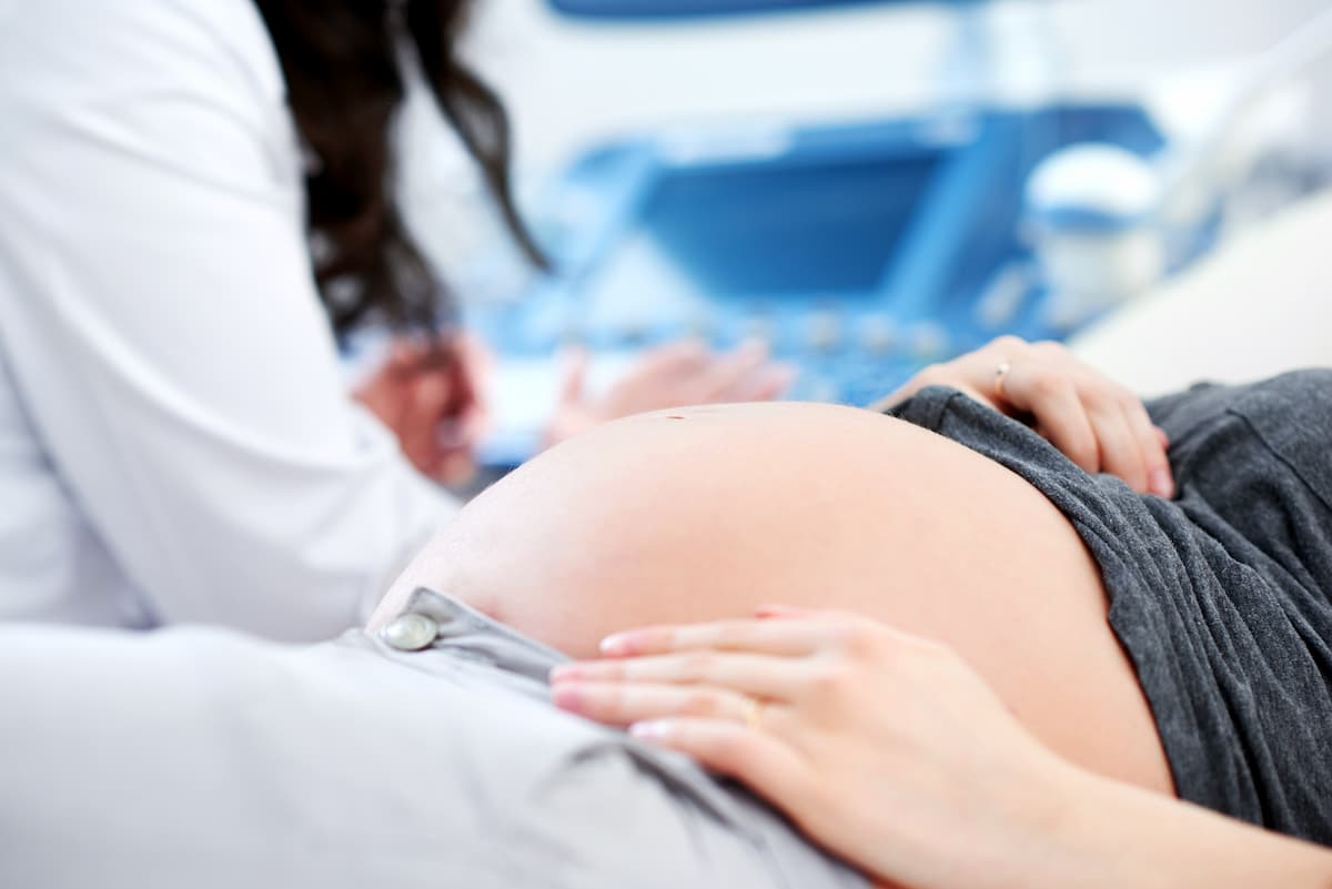
During pregnancy you will repeatedly hear the word "screening" from your doctor. This word sometimes causes anxiety, as it is associated with searching for problems. However, it is important to understand the true essence of this examination. Prenatal screening is not a diagnosis, but a special set of tests whose purpose is to assess the individual probability (risk) of certain fetal pathologies. It is a kind of "filter" that allows to identify a group of women who may need more careful monitoring or additional diagnostics. Undergoing screenings is a recommended and extremely important part of prenatal care.
Main purpose of screenings
The primary task of prenatal screenings is to assess the risk of the most common chromosomal abnormalities in the fetus, such as:
- Trisomy 21 (Down syndrome)
- Trisomy 18 (Edwards syndrome)
- Trisomy 13 (Patau syndrome)
In addition, screening ultrasounds allow assessment of overall child development and detection of possible organ and system malformations. For more details on why this is important, read the article "Genetic risks and screenings".
Combined screening first trimester (11 weeks – 13 weeks + 6 days)
This is the most important and most informative screening throughout pregnancy. It is called "combined" because it includes two tests whose results are evaluated together.
-
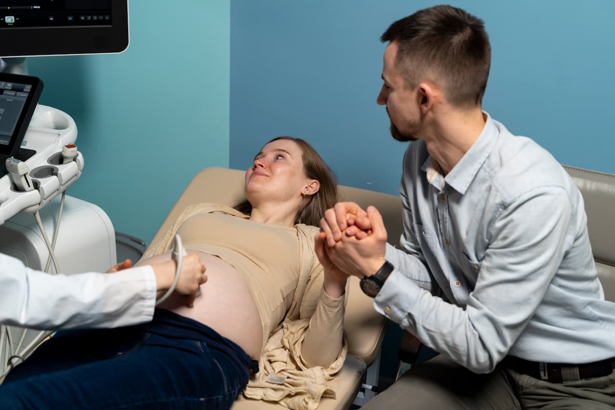
Ultrasound examination (US)
At this stage, ultrasound is not just the opportunity to see your baby for the first time, but also an important diagnostic procedure. The doctor carefully assesses:
- Gestational age: determined by fetal size.
- Nuchal translucency thickness (NT): This is a key marker. A small amount of fluid at the back of the fetal neck is measured. An increase in this measurement may indicate an elevated risk of chromosomal abnormalities.
- Presence of nasal bone: Its visualization is a good sign.
- Overall fetal anatomy: Main structures of the brain, limbs, abdominal wall are assessed.
-
Maternal blood biochemical analysis ("double test")
A blood sample is taken from the woman's vein to determine the levels of two specific proteins produced by the placenta:
- PAPP-A (pregnancy-associated plasma protein A)
- β-hCG (free β subunit of chorionic gonadotropin)
Deviations of these protein levels from the norm can also indicate increased risks.
Risk calculation: Ultrasound data, blood test results, as well as your age, weight and other individual parameters are entered into a special computer program (e.g., PRISCA), which calculates an individual risk as a ratio (e.g., 1:5000). The risk is considered low if the value is below the threshold (usually 1:250 or 1:300).
Second‑trimester screening (16-20 weeks)
This test is usually ordered if a woman, for some reason, missed the first‑trimester screening. It includes a biochemical blood analysis ("triple" or "quadruple" test), but is less accurate than the combined first‑trimester screening. The main event of the second trimester is a detailed ultrasound at 18-22 weeks, during which the doctor carefully examines all the baby's organs.
Non‑invasive prenatal test (NIPT)
NIPT is a modern and highly accurate screening method that can be performed from the 10th week. Fetal DNA is isolated from the mother's blood, allowing assessment of the risk of the most common chromosomal abnormalities with over 99% accuracy. This test is not part of the standard protocol in Ukraine and is paid for, but may be recommended by a doctor in case of high risk based on combined screening results or at the woman's request.
What to do if the risk is high?
A high risk based on screening results is **not a diagnosis**. It is only an indication for further consultations and possibly diagnostic procedures. A geneticist may propose invasive diagnostics (chorionic villus sampling or amniocentesis), which alone can confirm or rule out the diagnosis with 100% certainty.
Remember that undergoing screenings is your voluntary decision. Their purpose is to provide you with complete information to make informed decisions about managing your pregnancy.